| |
|
|
| HomeExpertise Aesthetic / Cosmetic Surgery (Indoor admission basis) Diabetic Foot Surgery |
 |
Diabetic Foot Surgery |
| |

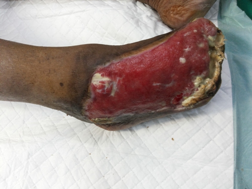
Diabetic Foot Surgery Before
|
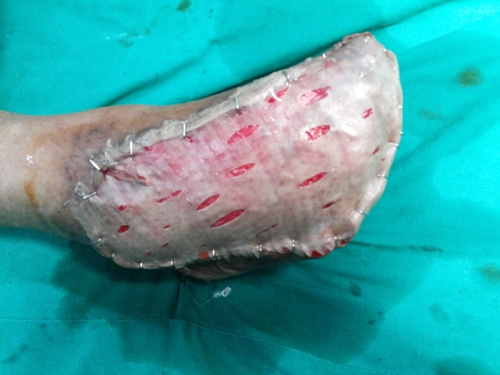
 Diabetic Foot Surgery After
|
|
Diabetic foot ulcers (DFUs) precede 85% of nontraumatic lower-extremity amputations (LEAs). Approximately 3-4% of individuals with diabetes currently have foot ulcers or deep infections. Fifteen percent develop foot ulcers during their lifetimes. Their risk of LEA increases by a factor of 8 once an ulcer develops. At 2 years following transtibial amputation, 36% have died.
Etiology : Peripheral neuropathy affects sensory, motor, and autonomic pathways
Sensory neuropathy : Sensory neuropathy deprives the patient of early warning signs of pain or pressure from footwear, from inadequate soft tissue padding, or from infection. This neuropathy appears in a stocking-glove distribution, with many of these individuals complaining of burning or searing pain.
- The primary risk factor for the development of DFUs is loss of protective sensation, best measured by insensitivity to the Semmes-Weinstein 5.07 (10 g) monofilament .
- Autonomic neuropathy produces chronic venous swelling. They lose normal vascular tone and thermal regulation, often developing severe venous swelling,also at times dry cracked skin due to loss of autonomic control.
- Motor peripheral neuropathy or Charcot osteoarthropathy can produce bony deformity, which, combined with the loss of protective sensation, can produce skin ulceration from pressure or from shear forces. Associated factors are history of foot infection or ulceration and previous partial or whole-foot amputation.
Ischemic peripheral vascular disease is the second risk factor for developing diabetic foot ulcer and infection. This used to be considered a small vessel disease, but current research links the vascular pathology to the basement membrane of the arterial wall. The disease is similar to that in those with vascular disease who are not diabetic, except that the distribution is somewhat more scattered and geographic in persons who are not diabetic, as opposed to progressive in a distal direction in persons who are diabetic.
The third major risk factor is related to the immune deficiency seen in this patient population. Glycosylated immune proteins lose efficiency, and granulocytes do not perform adequately, leaving these patients prone to infection from organisms that would not affect a healthy host.
Each of these potential abnormalities make the diabetic foot susceptible to abnormal mechanical stresses that can lead to a break in the normal soft tissue envelope, which can initiate a foot infection that cannot be resolved easily.
Pathophysiology:
- Pressure over bony prominence
- Skin breakdown
- loss of protective sensation
- leads to blister formation and full-thickness skin loss
- presence of severe venous swelling
- wound healing impaired by abnormally functioning white blood cells
|
| |
| |
|
| |
|
|